IMAT instrument scientist Dr. Genoveva Burca and her collaborators, who are research scientists from different universities, have published new research outlining the life science experiments that have taken place on IMAT and the growing capabilities of this instrument. From the initial mathematical modelling of the instrument to fine tuning techniques for life science applications, the scientists at ISIS have pioneered this new research on IMAT leaving no stone unturned to establish a reliable and distinctive form of analysis for biological samples. IMAT will be the first instrument that can do both neutron imaging and neutron diffraction, with hopes to bring together experts from a variety of disciplines to conduct non-destructive biological research.
Since 2016 IMAT has undertaken many cultural heritage and some engineering studies, but has now proved its relevance in the biological sector. These types of samples can often be fragile meaning neutron imaging analysis is ideal as it will not compromise or damage the sample, being entirely non-invasive. Most of the samples from the life sciences field are largely composed of hydrogen, making them especially suitable for neutron analysis. Where X-ray tomography can struggle to provide contrast, neutron imaging is especially good at detecting different isotopes of hydrogen. Neutrons attenuate differently to each isotope, shedding light on even subtle features of a sample. Experiments can be tailored for example, using deuterium, to highlight different parts of a sample uncovering structures previously unseen with other techniques, and this is continuously improving with the advancements to sources and detectors.
Paleontology
The delicate and rare nature of fossils makes them more challenging to image as it is essential to avoid damaging the sample. In recent decades, X-ray computed tomography (CT) scanning has transformed the way we approach and study fossils that are preserved in three dimensions. CT allows us to see inside these records of ancient life, and extract them digitally from their host rock. Some fossils, however, can't be studied in this way because X-rays are not able to distinguish between the fossil and the rock. When this is the case, paleontologists either can't study the fossils, or are forced to use destructive sampling techniques. The first fossil experiments on IMAT looked at 40 million year old fossilized crabs and a 160 million year old fossil ammonite. Led by a group of researchers from Oxford University, Manchester University, National History Museum and Imperial College London, the experiments showed that neutron tomography can really improve the paleontological research, because differences in composition which are invisible to X-rays were imaged using neutrons. Because fossils are made of many different minerals, having another tool palaeontologists can use to study them is a really exciting development. Therefore neutron tomography can have a strong impact in the field of paleontology and give insights into the relationships between different species and their evolutionary history.
Credit: Dr I A Rahman – Oxford University, OUMNH, Dr R J Garwood – Manchester University, NHM and Dr A.R.T. Spencer – Imperial College London, NHM, Dr G Burca – ISIS Facility
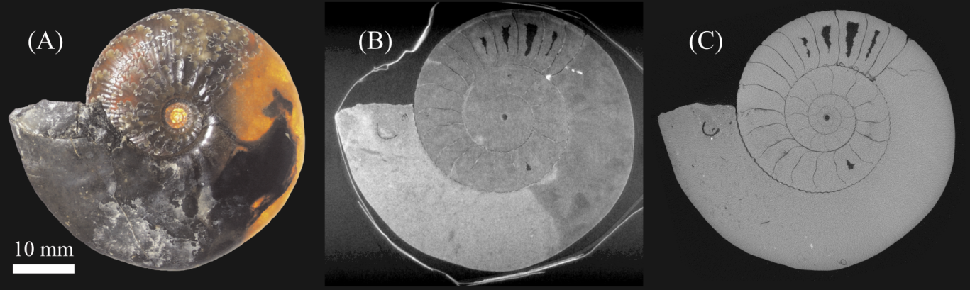
Figure 1. Exceptionally preserved three-dimensional fossil ammonite from the Jurassic (~166 million years ago) of England. (A) Photograph of fossil specimen. (B) Slice from neutron tomography scan at IMAT of fossil shown in (A). (C) Slice from X-ray computed tomography scan of fossil shown in (A).
Soil-plant science
Moving away from fossils and into the present day, the global challenges of increased water restraints on the agricultural industry and food security issues has prompted soil-plant studies. X-ray imaging gives clear insights into soil structure and composition, however water is comparatively transparent to X-rays and biological matter also shows poor contrast. Neutron imaging presents a complementary probe where water and biological matter are better distinguished but the soil minerals are not imaged as clearly as they would be with X-rays. This research aims to develop robust methods for complementary X-ray/neutron tomographic imaging of plant-root systems to gain new insights into water transport and nutrient uptake in soil. The first soil measurement on IMAT found that compost is effective at improving water and nutrient distribution as it can absorb more than its own weight in water. The study also showed that water is predominately held within clay rather than grains of minerals within the soil. And this is just a small part of the agricultural potential of IMAT, where drought resistant plants could also be studied to combat global problems.
Credit: Mr T Clark, Dr T Blumensath, Dr R Boardman, Dr S Keyes (Southampton University), Dr G Burca – ISIS Facility
Figure 2. A preliminary dataset of a lupin seedling in soil, grown in a glass sample tube (boron free) with cadmium fiducial markers attached externally and serves to demonstrate the complementarity of the two modalities (X-ray and neutron imaging). The middle of the three circles shows the two techniques combined, within this, the round green shape represents the seedling, the other green areas show water distribution, and the red parts are soil and minerals.
Dentistry
After 1049 image projections collected over 9 hours measurement, the first neutron tomography of a human tooth was completed on IMAT. The internal structure was revealed exposing the pulp chamber, dentin and enamel layers. The 3D image of a second molar was optimized using normalization to eliminate background 'noise' in the image. This kind of imaging could have applications in endodontic processes such as dental pulp treatment but also in forensic science, demonstrating how the capabilities of IMAT could have a significant impact in a range of research fields related to public health.
Figure 3. 3D images of a second molar with the pulp chamber (red), dentin (green) and enamel (white).
This research demonstrates the array of samples that can be analysed with this technology and the fascinating results that can be obtained using this complementary technique. With the number of life science proposals already on the rise for the instrument, it is evident that the amount of exciting new science stemming from IMAT is only going to increase. This recent work highlights the unique capabilities and potential of IMAT that will have a significant impact on life science innovation and we are eagerly awaiting its next triumph.
The full publication is available to view in the Journal of Microscopy, The Royal Microscopical Society.
For further information about the research, please contact Dr. Genoveva Burca (Genoveva.Burca@stfc.ac.uk)
Browse all our science highlights here.
Find further information on IMAT