The root system is key to plant functions such as water and nutrient uptake, anchorage and symbiotic interactions, with processes occurring at the root-soil interface strongly controlling plant performance. Therefore, studies into root systems and their interactions with the soil matrix could provide valuable insight into mechanisms that maximize crop yield. Crops with improved traits below the ground could lead to enhanced plant performance and an increase in crop yield, helping to address global food security challenges and meet the demands of a growing population.
Tomographic imaging is ideal for plant root studies since it is non-invasive and allows for in vivo investigations. However, no single imaging technique is well suited to capture the whole system. While X-ray imaging gives insight into soil composition and structure, there is very little contrast between water, plant roots and any other biological soil constituents. Neutrons here offer a complimentary view where water and biological matter are clearly distinguished, allowing water dynamics around the roots to be investigated. It is the combination of neutrons and X-rays that is needed to capture the complete system and provide a clearer understanding of root-soil interactions.
In order to align the datasets from the two different modes of imaging, an object or marker can be attached to a sample as a point of reference. These are known as fiducial markers. For 3D images, at least three fiducial points must be identified in both the reference and target images so that the parameters for alignment can be found and a suitable transform applied. In their most recent paper, the researchers investigated the use of cadmium fiducial markers.

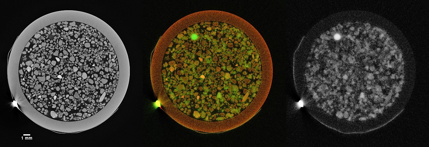
Figure 1. A slice from a plant root sample with X-ray data on the left and in red in the middle image, and neutron data on the right and in green in the middle image. This slice shows the match of a fiducial marker and the complementarity of the different modes of imaging (Clark et al., 2019).
The experiment using the cadmium markers demonstrated that more information can be collected by combining imaging techniques than either technique, X-ray or neutron, in isolation (Figure 1). However, cadmium's high attenuation was found to introduce artefacts in the images that were shown to have a negative impact on data alignment. While cadmium is not an ideal fiducial marker, this work clearly demonstrates the value of combining neutron and X-ray tomography for studying plant root systems. The team will be back at ISIS and Diamond to investigate the suitability of other materials as fiducial markers to provide new insights into water and nutrient transport in plants and soil.
Further information
More information on correlative neutron and X-ray imaging of root systems can be found in T. Clark et al. (2019), Journal of Microscopy, https://doi.org/10.1111/jmi.12831.
More examples of bioscience research on IMAT.
